5
D
:
5
H
:
5
M
:
5
S
from sept 25 to 27, 2024

Symposium 1

New imaging techniques and innovative approaches

WEDNESDAY, SEPTEMBER 25, 2:15 p.m.

#1
Meera Srikrishna

CT-based imaging markers for idiopathic normal pressure hydrocephalus obtained using deep learning: association with MRI-based radiological markers and diagnosis

Background: Brain computed tomography (CT) is a widely accessible and affordable modality routinely used to assess neuroimaging signatures of idiopathic normal pressure hydrocephalus (iNPH), particularly ventricular enlargement. Previously, we developed deep learning models to segment various brain tissue classes in brain CTs. This study aims to develop novel tools capable of automatically extracting volumetric measurements and ratios from CT brain scans. Further, we aim to assess the association of CT-based volumetric measures (CTVMs) with radiological markers of iNPH and diagnosis.

Methods: We included 777 (52% female, 70.44 ± 2.6 years) CT scans which had paired T1-weighted magnetic resonance image scans from the Gothenburg H70 Birth Cohort studies collected on the same day. An experienced rater evaluated various iNPH-related radiological markers in all T1-weighted images. Thirty-nine participants fulfilled the criteria of radiological probable iNPH.  Our trained U-Net-based deep learning models were used to predict brain volume (BV; obtained by the summation of grey matter (GM) and white matter (WM) tissue classes), intracranial volume (ICV), cerebrospinal fluid (CSF), and ventricular CSF (VCSF) segmentations from input CT scans.  CT-based volumetric measures (CTVMs) such as VCSF/ICV, BV/ICV, and VCSF/CSF were derived. The relationship between CTVMs and standard radiological markers such as Evan’s index, Z-Evan’s index, and callosal angle was assessed using Spearman’s rank correlation tests.

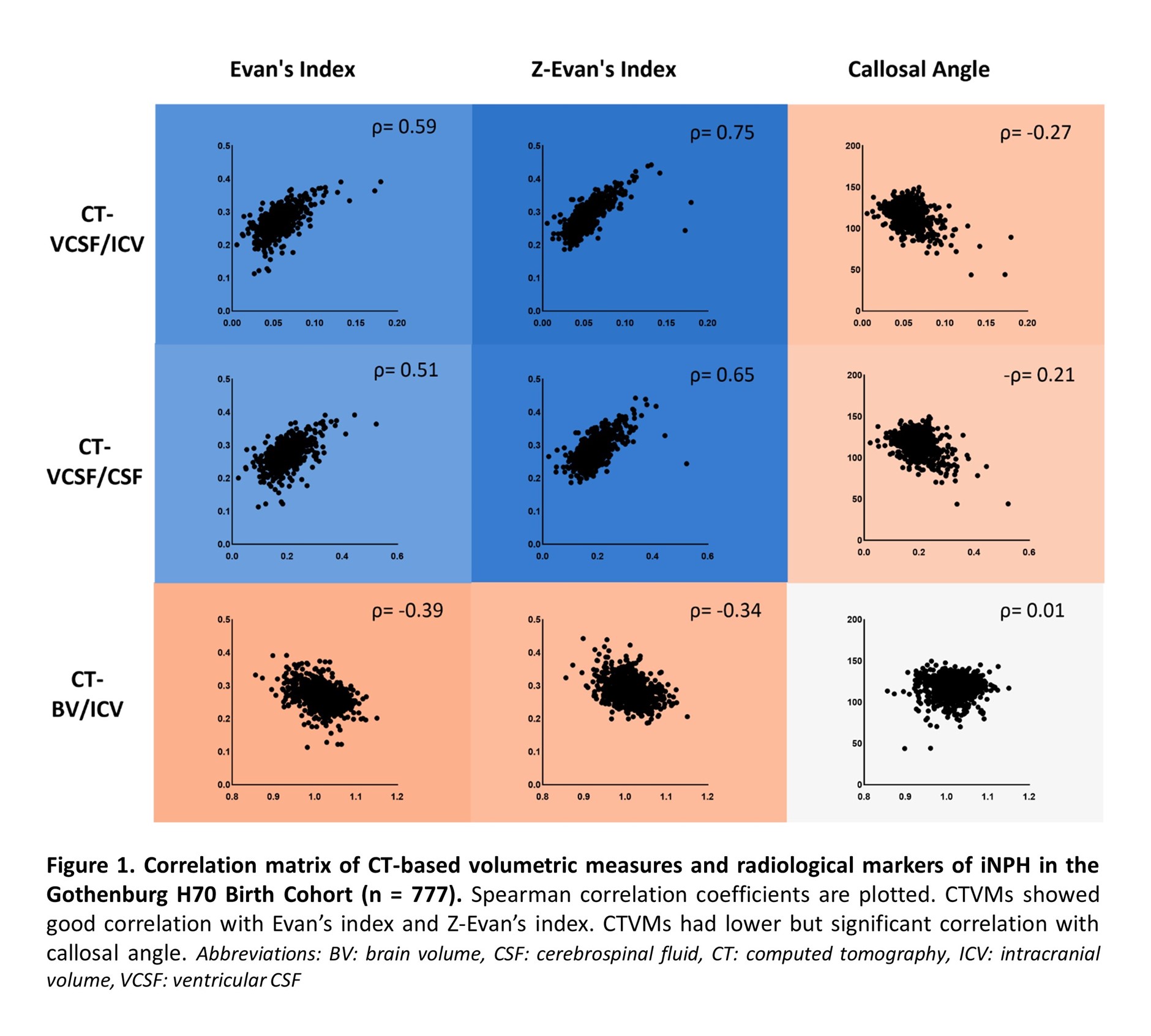
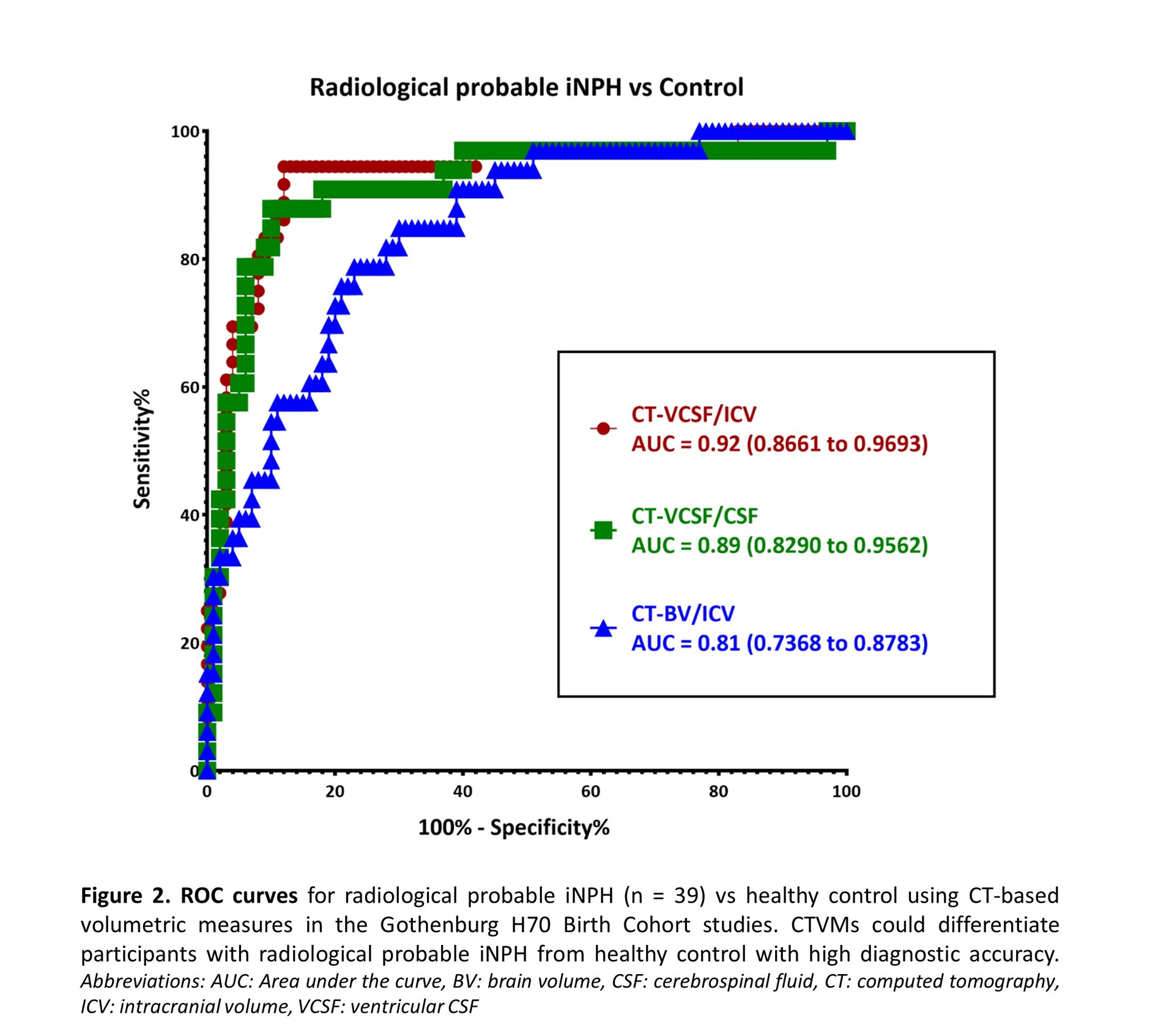
Results: CT-based volumetric measures could differentiate participants with radiological probable iNPH from healthy control with high diagnostic accuracy (Area under the curve: 0.92 for CT-VCSF/ICV). CTVMs showed good correlation with Evan’s index (ρ=0.59, p<0.001) and Z-Evan’s index (ρ=0.75, p<0.001). CTVMs had a lower but significant correlation with callosal angle (ρ=-0.27, p<0.001). CT-VCSF/ICV showed a higher correlation with radiological markers of iNPH in comparison to CT-BV/ICV and CT-VCSF/CSF. Higher levels of Evan’s index and Z-index are associated with higher levels of CT-VCSF/ICV and CT-VCSF/CSF and lower levels of CT-BV/ICV.

Conclusion: CT-based volumetric measures correlate with relevant radiological markers of iNPH. CTVMs could distinguish radiological probable iNPH and controls. This supports the potential application of CTVMs in aiding diagnostics and monitoring the progression of iNPH. Automated CT-based angular measurements and the association of CTVMs with other clinical iNPH markers will be further explored in future.

Authors: Meera Srikrishna1,2,3, Clara Constantinescu4,5, Doerthe Ziegelitz6, Anna Zettergren7, Silke Kern7,8, Lars-Olof Wahlund9, Eric Westman9, Ingmar Skoog7, Mats Tullberg4, Michael Schöll1,2,3,10,11

Author Affiliations:

  1. Centile Bioscience, USA & UK;
  2. Wallenberg Centre for Molecular and Translational Medicine, University of Gothenburg, Sweden;
  3. Department of Psychiatry and Neurochemistry, Institute of Physiology and Neuroscience, University of Gothenburg, Sweden;
  4. Department of Clinical Neuroscience, Institute of Neuroscience and Physiology, The Sahlgrenska Academy, University of Gothenburg, Sahlgrenska University Hospital, Sweden;
  5. Department of Neurology, Sahlgrenska University Hospital, Sweden;
  6. Department of Radiology, Institute of Clinical Sciences, Sahlgrenska Academy, University of Gothenburg, Sahlgrenska University Hospital;
  7. Neuropsychiatric Epidemiology, Institute of Neuroscience and Physiology, Sahlgrenska Academy, Centre for Ageing and Health (AgeCap), University of Gothenburg, Sweden;
  8. Department of Psychiatry and Neurochemistry, Institute of Neuroscience and Physiology, Sahlgrenska Academy, University of Gothenburg, Mölndal, Sweden;
  9. Division of Clinical Geriatrics, Department of Neurobiology, Care Sciences and Society, Karolinska Institutet, Sweden;
  10. 1Dementia Research Centre, Institute of Neurology, University College London, UK;
  11. Department of Clinical Physiology, Sahlgrenska University Hospital, Gothenburg, Sweden

#2
Heidi Jacobs

Locus coeruleus imaging to identify pre-preclinical Alzheimer’s disease


Background: Alzheimer’s disease (AD) is characterized by a long asymptomatic stage during which the two pathologic hallmarks, beta-amyloid and tau depositions, accumulate, lead to neurodegeneration and the manifestation of clinical symptoms. Being able to effectively delay disease progression will require early intervention, before irreversible damage has occurred. There is thus need for a biomarker that can predict who is likely to accumulate AD pathology and show a decrease in cognition in the following years. Given that autopsy data reported presence of hyperphosphorylated tau in the locus coeruleus (LC) early in adulthood and prior to cortical involvement, we aimed to evaluate the LC using structural neuroimaging as a promising early marker of AD risk.


Methods

We used two different cohorts: 1) data from Harvard Aging Brain Study+ (n=214) where individuals underwent 3T MRI-LC imaging, 18F-Flortaucipir (FTP)-PET imaging, PiB-PET imaging and longitudinal cognitive assessments. Of these individuals, 77 underwent repeated LC imaging and repeated FTP-PET imaging; 2) data from the adult lifespan study (n=99) where individuals underwent 7T MRI-LC imaging and cognitive assessments and of whom novel plasma markers were quantified (ptau181, ptau217, ptau231 and Aß42/40)

Results: Lower LC integrity was associated with greater entorhinal tau and PiB-related cognitive decline (starting earlier than the established PiB threshold). Longitudinal data revealed that lower LC integrity preceded accumulation of tau in the medial temporal lobe, and that the relationship between lower baseline LC integrity and lower follow-up cognitive performance was mediated by accumulation of medial temporal lobe tau. When comparing LC integrity, PiB and hippocampal volume in all PiB- individuals, LC integrity was the best predictor of entorhinal tau accumulation and disease progression. In the second dataset, we observed that lower dorso-rostral LC integrity was associated with greater ptau231 and ptau217, starting from age 54 years, and together both events predicted lower cognitive performance. We found no relationships between LC integrity and plasma Aß42/40.

Conclusions: Our findings highlight that LC changes reflect early pathologic changes relevant to AD, predict disease progression and identify LC imaging as a promising indicator of initial AD-related processes.

Keywords: Locus coeruleus, early detection, neuroimaging, plasma markers, ultra-high field imaging

Authors:

  • Heidi, Jacobs, The Athinoula A. Martinos Center for Biomedical Imaging, Department of Radiology, Massachusetts General Hospital / Harvard Medical School, Boston
  • John Alex, Becker, Gordon Center for Medical Imaging, Department of Radiology, Massachusetts General Hospital / Harvard Medical School, Boston
  • Nina, Engels-Domínguez, The Athinoula A. Martinos Center for Biomedical Imaging, Department of Radiology, Massachusetts General Hospital / Harvard Medical School, Boston
  • Nicholas, Ashton, Department of Psychiatry and Neurochemistry, Institute of Neuroscience and Physiology, The Sahlgrenska Academy at the University of Gothenburg, Mölndal, Sweden
  • Oskar, Hansson, Clinical Memory Research Unit, Department of Clinical Sciences Malmö, Lund University, Lund, Sweden
  • Henrik, Zetterberg, Department of Psychiatry and Neurochemistry, Institute of Neuroscience and Physiology, The Sahlgrenska Academy at the University of Gothenburg, Mölndal, Sweden
  • Prokopis, Prokopiou, The Athinoula A. Martinos Center for Biomedical Imaging, Department of Radiology, Massachusetts General Hospital / Harvard Medical School, Boston
  • Dorene, Rentz, Center for Alzheimer Research and Treatment, Department of Neurology, Brigham and Women’s Hospital, Boston
  • Julie, Price, The Athinoula A. Martinos Center for Biomedical Imaging, Department of Radiology, Massachusetts General Hospital / Harvard Medical School, Boston
  • Reisa, Sperling, Center for Alzheimer Research and Treatment, Department of Neurology, Brigham and Women’s Hospital, Boston
  • Keith, Johnson, Gordon Center for Medical Imaging, Department of Radiology, Massachusetts General Hospital / Harvard Medical School, Boston

#3
Mitsuko Nakajima

Differential cortical layer vulnerability in premanifest Huntington’s disease.


Background: Huntington’s Disease (HD), a dominantly inherited, fatal neurodegenerative disease is characterised by progressive striatal and cortical atrophy. Post-mortem studies in advanced HD have shown selective atrophy in layer 3 (L3) and L5 of the primary motor cortex, and L3, L4 and L6 of the primary visual cortex.1,2 However, the distribution and extent of cortical layer pathology in premanifest HD (preHD) is unknown.

Methods: Using ultra-high field (UHF) 7-Tesla MRI at 600µm isotropic resolution, we employed a whole brain voxel-wise analysis of cortical volumes and quantitative (qMRI) measures to compare preHD and healthy controls. Family-wise error-corrected voxels were overlaid with the Human Connectome Project Multi-Modal Parcellation (HCP-MMP) 1.0 atlas to identify target regions of interests (ROIs) for cortical layer analyses. The ROIs were then divided into 8 equi-volume layers, and qMRI measurements per layer were compared between preHD and controls. The von Economo (VE) atlas was used to estimate histological cortical layers.

Results: Volumetric analysis showed atrophy in the striatum but not in the cortex. Voxel-wise analysis revealed two clusters with lower Proton Density (PD) values in preHD (n=16) than controls (n=18) which were localised to left frontal eye field (FEF) and right visual area 6 (V6) of the HCP-MMP 1.0 atlas. In left FEF, significant differences were observed in equi-volume L1, L4, L7, and L8, with the largest effect (P=0.03, Hedges’ d=0.95) in L4. In right V6, differences were observed in equi-volume L2-L8, with the largest effect again in L4 (P=0.01, Hedges’ d=1.26). These regions were localised to left area frontalis agranularis (FB) and right area peristriata (OA) of the VE atlas. In left FB, histological layer III had the largest effect (P=0.009, Hedges’ d=1.15), and in right OA, layer V (P=0.0001, Hedges’ d=1.7).

Conclusions: This is the first study to demonstrate differential cortical layer vulnerability in preHD. Histological layer V showing the largest effect in right OA is consistent with previously reported post-mortem studies typically performed in advanced HD.3 We observed group differences in the cortex with qMRI but not with volumetric MRI, suggesting a novel role for UHF 7T qMRI in examining cortical pathology in preHD.

REFERENCES

  1. Thu DCV, Oorschot DE, Tippett LJ, Nana AL, Hogg VM, Synek BJ, et al. Cell loss in the motor and cingulate cortex correlates with symptomatology in Huntington’s disease. Brain. 2010;133(4):1094–110.
  2. Macdonald V, Halliday G. Pyramidal cell loss in motor cortices in Huntington’s disease. Neurobiol Dis. 2002;10(3).
  3. Pressl C, Mätlik K, Kus L, Darnell P, Luo JD, Weiss AR, et al. Layer 5a Corticostriatal Projection Neurons are Selectively Vulnerable in Huntington’s Disease [Internet]. bioRxiv; 2023 [cited 2023 Apr 26]. p. 2023.04.24.538096. Available from: https://www.biorxiv.org/content/10.1101/2023.04.24.538096v1

Tables or Figures:

Figure 1 Flowchart of analysis pipeline. MPM – multi-parametric map; VBQ – voxel-based quantification; ROI – region of interest; MNI – Montreal Neurological Institute.

Figure 2 Localising the regions of interest to von Economo histological atlas. The two significant clusters from group comparison from voxel-based quantification (VBQ, shown in red in A and B) were localised to identify the regions of interest from the von Economo histological atlas (yellow in A and B). The clusters were localised to the left area frontalis agranularis (FB) (A) and the right area peristriata (OA)(B). Each ROI was divided into eight equi-volume layers using Nighres. (C) shows results for left FB , and (D) shows results for right OA.

Figure 3 Equi-volume layer results per von Economo ROIs with estimated histological layer. Boxplots representing median and IQR per group for each equi-volume layer for left FB and right OA. Approximated histological layers based on von Economo atlas are shown as coloured bars. Keys: c – controls; p – preHD; * = p value <0.05; ** = p value <0.01; *** = p value <0.001. Abbrev: FB – area frontalis agranularis; OA – area peristriata; PD – proton density; GM – grey matter; WM – white matter.

Keywords: Huntington’s Disease; ultra-high field MRI; cortical layers; multiparametric mapping; quantitative MRI

Authors and affiliation:

  • Mitsuko Nakajima1
  • Rachael I Scahill1
  • Nicola Z Hobbs1
  • Martina F Callaghan2
  • Kate Fayer1
  • Geraint Rees3
  • Sarah J Tabrizi1
  • Peter McColgan1

Affiliation:
1 Huntington’s Disease Centre, Department of Neurodegenerative disease, UCL Queen Square Institute of Neurology, University College London, London, UK.

2 Wellcome Centre for Human Neuroimaging, Department of Imaging Neuroscience, UCL Queen Square Institute of Neurology, University College London, London, UK.

3 Institute of Cognitive Neuroscience, University College London, London, UK

= These authors contributed equally to this work and share senior authorship

#4
Joana Pereira

Understanding the role of the neuromodulatory nuclei in brain connectivity in aging and neurodegeneration?


Background: The communication between cortical and subcortical areas, also known as brain connectivity, is a reliable biomarker of downstream cognitive impairment and upstream neuronal alterations, being profoundly influenced by the neuromodulatory nuclei. These nuclei, including the locus coeruleus and substantia nigra, play a crucial role in the release and regulation of neurotransmitters across the brain, modulating various cognitive processes such as attention, learning, and memory, as well as emotional and behavioral responses.

Methods: In this series of studies, we utilized a gradient-based approach to evaluate the functional and anatomical connections of the locus coeruleus and substantia nigra across a lifespan cohort of cognitively normal individuals in addition to patients at varying stages of Alzheimer’s and Parkinson’s diseases. We explored the association between connectivity gradients and various outcomes, including cognition, psychiatric symptoms, motor function, sleep, and other non-motor measures, through diverse statistical analyses.

Results: Our findings indicate that the functional and anatomical connectivity of the rostral and dorsal parts of the locus coeruleus are particularly susceptible to aging, mirroring early alterations observed in Alzheimer’s disease. This vulnerability is linked to worse emotional memory, language, visuoperception, and sleep quality. Moreover, a pattern of reduced connectivity from the substantia nigra to motor and sensorimotor regions was observed with advancing age, correlating with deficits in motor learning, working memory, and Parkinson’s disease.

Conclusions: The results of these studies underscore the critical role that neuromodulatory nuclei play in brain connectivity. They suggest that strategies aimed at preserving or augmenting neuromodulatory functions may offer promising therapeutic targets for attenuating cognitive and motor decline related to aging and neurodegenerative conditions such as Alzheimer’s and Parkinson’s diseases.


NEWSLETTER

This website uses cookies to ensure audience measurement, as well as sharing or redirection to social networks. (Know more)
OK