5
D
:
5
H
:
5
M
:
5
S
from sept 25 to 27, 2024

Symposium 3

Opportunities and Challenges in Data Sharing in European Neuroimaging

THURSDAY, SEPTEMBER 26, 10:30 a.m.

#10
Julia Neltzel

Data sharing strategies in the Rotterdam Study


Abstract: I will first give a brief overview of the prospective, population-based Rotterdam Study and highlight some key recent findings of our brain imaging research lines. In one recent development, we have enriched a sub-sample with amyloid PET and state-of-the-art AD plasma markers, allowing us to investigate the complex interplay between vascular and AD pathology.

I will dedicate the larger part of the talk to our experience with data sharing. While data sharing is a key element of science today, it wasn’t when the Rotterdam Study started in 1989. For this reason, our participants’ informed consent form still contains some restrictions on how data can be shared outside our research centre. I will present strategies of how we nevertheless shared data with external collaborators, large consortia, and with the public. According to a review by Mills & Rahal (2019), the Rotterdam Study was the most frequently utilized data set across the largest GWAS. Meta-analysis is an approach to sharing summary statistics that is often used in genetics, but can also be applied to brain imaging. I will present an unpublished example in which we meta-analysed results of six large cohort studies from the Cross-Cohort Collaboration consortium (CCC) on the associations of education, marital status, and physical activity with brain health markers. Yet, some techniques, like machine learning, require the actual data. To get around the need to share data centrally, we have started to develop federated learning platforms, that allow models to be trained on multiple decentralised servers with local data samples. Another effort is to share data with the public. Together with ten partners from three European countries, we created the JoinUs4Health platform with the mission to engage study participants, and any interested citizen in health science. This secure online environment allows anyone to work as or with a scientist on pressing health issues. The Rotterdam Study is one of three cohort studies that can be accessed via this platform.

I will conclude my presentation by discussing some of the challenges that remain and looking ahead to the developments on the horizon.

Keywords: Epidemiology, brain imaging, data sharing, meta-analysis, federated learning

Author: Julia, Neitzel, Department of Radiology & Nuclear Medicine, Department of Epidemiology, Erasmus MC University Medical Center Rotterdam, The Netherlands Department of Epidemiology, Harvard TH Chan School of Public Health, Boston, United States

#11
Oliver Speck

Opportunities and Challenges in multicenter
MR neuroimaging studies


Abstract: Multicenter imaging trials open the path to large and distributed cohort studies. The inclusion of multiple centers, however, poses significant challenges with respect to harmonized image data acquisition, meta data handling, data transfer and data processing as well as data privacy issues. Within the DELCODE study, a large multicenter trial including MR imaging in subjects with mild cognitive impairment, more than 5000 imaging sessions have been acquired in 11 centers since 2014. After an initial consensus process on the imaging protocols, the detailed implementation on different scanners and the maintenance with scanner upgrades is one challenge. Training of and constant feedback to MR operators has been identified as a key aspect to high data quality that reduced low quality data over the initial phases of the study. With long term follow ups and study continuation, protocol updates are required to take advantage of modern acquisition techniques. Major efforts in protocol piloting are required to ensure data continuity. As a result, a unique and valuable dataset that already served as the basis for many high-level publications is available and continues to be expanded.

Keywords: multicenter studies, harmonization, data quality management

Authors:

  • Oliver Speck, Otto-von-Guericke University Magdeburg, DZNE Magdeburg
  • Emrah Düzel, DZNE Magdeburg
  • Falk Lüsebrink, DZNE Magdeburg

#12
Ludovica Griffanti

From ‘big data’ to the clinic: implementing the UK Biobank imaging framework for memory clinic patients

Background: UK Biobank (UKB) is a large cohort study with 500,000 participants, 100,000 of which are undergoing detailed brain MRI. Projects like UKB led to the development of image analysis and statistical methods capable of handling big datasets, providing the power to reveal novel associations between imaging derived phenotypes (IDPs) and non-imaging variables. I will summarise some example studies, highlighting how this shared resource can inform dementia research. While the large size of the UKB dataset is vital for these sorts of studies, both the ascertainment of dementia diagnosis and dementia-specific phenotyping are inevitably limited given the scale of the data. Smaller research cohorts designed for dementia tend to be highly selected and poorly representative of patients (particularly the majority who present to psychiatry-based memory services in the UK), and there is a disconnect between the methods used in research and clinical settings.

Methods: To address the gap between research and clinic, we developed infrastructure, the Oxford Brain Health Clinic (OBHC), to integrate research assessments into the clinical pathway for memory clinic patients, including incorporating a brain MRI protocol matched with UKB. We then adapted the UKB image analysis pipeline to be more robust on patients’ data and to include IDPs intended to capture dementia-related changes. To demonstrate the usability of the OBHC dataset, we tested associations between IDPs and cognition using linear regression on data from 201 patients. Finally, we implemented statistical adaptations (hierarchical FDR adjustment) to enable big data-style analyses in this representative memory clinic sample.

Results: Linear regression analyses validated known associations between cognitive performance and IDPs measuring cortical atrophy, vascular pathology and white matter integrity in a real-world memory clinic population. We also observed different patterns of associations between IDPs and cognitive domains. For example, memory showed mostly associations with structural IDPs (Figure 1A) while the visuospatial domain included associations with functional IDPs (Figure 1B).

Conclusion: The alignment of data acquired within routine clinical care with the UKB gives us a unique opportunity to translate models developed using ‘big data’ into a real-world population. Ultimately, we hope to produce evidence to improve differential diagnosis and treatment pathway decisions for patients.

Figure:

Figure 1. Manhattan plot of associations between IDPs and memory (A) or visuospatial skills (B). The hight of each dot represents the strength of the association between one IDP and the memory (A) or visuospatial (B) domain score of the Addenbrooke’s Cognitive Examination III (ACE III). IDPs are grouped along the x axis according to the different types of scans and analysis tools used to calculate them, and they are color-coded by MRI modality. Correction for multiple comparisons was performed using hierarchical FDR adjustment, first across modalities then within modality (black horizontal line represents the FDR-corrected threshold).

Keywords: MRI, UK Biobank, Big data, Dementia, Clinical translation

Authors:

  • Ludovica Griffanti, University of Oxford
  • Grace Gillis, University of Oxford
  • Gaurav Bhalerao, University of Oxford
  • Jasmine Blane, University of Oxford
  • Robert Mitchell, Oxford Health NHS Foundation Trust
  • Celeste McCracken, University of Oxford
  • Thomas Nichols, University of Oxford
  • Lola Martos, Oxford Health NHS Foundation Trust
  • Vanessa Raymont, University of Oxford
  • Clare Mackay, University of Oxford

#13
Lyduine Collij

From AMYPAD to EuroPAD – creating a multi-cohort preclinical database to model AD disease trajectorie


Background: The emergence of disease-modifying drug therapies is expected to revolutionize the field of Alzheimer’s disease (AD). Recent results from anti-amyloid clinical trials highlight the importance of early identification and accurate risk-stratification of individuals in early stages of the disease. In this context, the Amyloid Imaging to Prevent Alzheimer’s Disease (AMYPAD) Prognostic and Natural History Study (PNHS) was established, leveraging existing cohorts to alleviate the burden of recruiting de novo participants1, 2. Here, we describe the harmonization and integration efforts of imaging, clinical, cognitive, and fluid biomarker data.

Access to the data is available through the Alzheimer’s Disease Data Initiative, with additional details provided at https://amypad.eu/data/

Methods: The AMYPAD-PNHS integrates prospective and historical data from 32 European sites across 10 countries currently3. These sites contribute data from 10 Parent Cohorts (PC), predominantly comprising non-demented at-risk subjects, including EPAD LCS, EMIF-AD (60++ and 90+), ALFA+, FACEHBI, FPACK, UCL-2010-412, Microbiota, DELCODE, and the AMYPAD Diagnostic and Patient Management Study (DPMS).

A meticulous data curation process was implemented, harmonizing metrics and questionnaires through strategies such as recoding into categories, Percentage of Maximum Possible Scores, and z-scores. Expert reviewers at each site conducted PET visual reads. Centralized quantification of static PET images, employing site-specific Gaussian smoothing, yielded harmonized Centiloid values. Parametric modelling of dynamic PET scans was also performed providing metrics such as the distribution volume ratio (DVR).

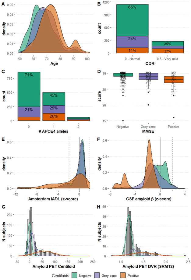
Results: The initial data set includes 3366 participants (55% females, 67±8 years), with 2629 having at least one follow-up visit (2.56±1.91 years). Of those, 1618 underwent baseline amyloid-PET, 888 with follow-up. The dataset incorporates clinical outcomes, biomarkers, risk factors, and other pertinent variables (Figure-1). Distribution of participants based on amyloid PET status at baseline is of 60% negative (<12CL), 24% grey-zone (12-50CL), and 16% positive (>50CL).

Conclusions: The AMYPAD-PNHS represents the largest European longitudinal dataset phenotyping individuals at risk of AD-related progression. The consortium is currently evolving into its new phase, namely the EuroPAD collaborative framework, and the dataset will be expanded in terms of variables (e.g., currently integrating GWAS and advanced MRI data) and additional cohorts.

Figure 1. Representative set of variables contained harmonized and integrated into the AMYPAD PNHS dataset.

Keywords: Alzheimer’s disease, Amyloid-PET, Magnetic Resonance Imaging, consortium, disease modelling

Authors affiliations: Lyduine E. Collij, PhD1,2, David Vállez García, PhD1, Willemijn J. Jansen, PhD3, Stephanie J. B. Vos, PhD3, Julie Elisabeth Oomens, MSc4, Natalia Vilor-Tejedor, PhD5, Laura Stankeviciute, PhD5, Marta Mila-Aloma, PhD5, Anna E Leeuwis, PhD1, Gonzalo Sánchez-Benavides, PhD5, Mahnaz Shekari, MSc6, Fiona Heeman, PhD7, Luigi Lorenzini, MSc1, Leonard Pieperhoff, MSc1, Bert Overduin, PhD8, Isadora Lopes Alves, PhD9, Juan Domingo Gispert, PhD5, Frank Jessen, MD10, Pieter Jelle Visser, MD, PhD1, Anouk den Braber, PhD1, Craig W Ritchie, MBChB, PhD11, Mercè Boada, MD, PhD12, Marta Marquié, MD, PhD13, Rik Vandenberghe, MD, PhD14, Emma S. Luckett, PhD15, Michael Schöll, PhD7, Giovanni B. Frisoni, PhD, MD16, Bernard J Hanseeuw, MD, PhD17, Lisa Quenon, PhD17, Andrew W. Stephens, MD, PhD18, Lisa Ford, MD19, Mark E Schmidt, MD20, Cindy Birck, PhD21, Anja Mett, BSc22, Rossella Gismondi, MD18, Richard Manber, PhD23, Sylke Grootoonk, PhD23, Robin Wolz, PhD24, Gill Farrar, PhD25, Frederik Barkhof, Prof1,26 on behalf of the AMYPAD Consortium

 (1)Amsterdam UMC, Amsterdam, Netherlands, (2)Lund University, Lund, Sweden, (3)Maastricht University, Maastricht, Netherlands, (4)Alzheimer Center Limburg, School for Mental Health and Neuroscience, Maastricht University, Maastricht, Netherlands, (5)Barcelonaβeta Brain Research Center (BBRC), Barcelona, Spain, (6)Barcelonaβeta Brain Research Center (BBRC), Pasqual Maragall Foundation, Barcelona, Spain, (7)University of Gothenburg, Gothenburg, Sweden, (8)Aridhia Informatics Ltd, Glasgow, United Kingdom, (9)Brain Research Center, Amsterdam, Netherlands, (10)German Center for Neurodegenerative Diseases (DZNE), Bonn, Germany, (11)Scottish Brain Sciences, Edinburgh, United Kingdom, (12)Ace Alzheimer Center, Barcelona, Spain, (13)Ace Alzheimer Center Barcelona, Barcelona, Spain, (14)University Hospitals Leuven, Leuven, Belgium, (15)Karolinska Institutet, Stockholm, Sweden, (16)University of Geneva, Geneva, Switzerland, (17)UC Louvain, Brussels, Belgium, (18)Life Molecular Imaging GmbH, Berlin, Germany, (19)Janssen Pharmaceutica, Titusville, NJ, USA, (20)Janssen Pharmaceutica, Beerse, Belgium, (21)Alzheimer Europe, Luxembourg, Luxembourg, (22)GE Healthcare, Amersham, United Kingdom, (23)IXICO, London, United Kingdom, (24)IXICO, London, Greater London, United Kingdom, (25)GE HealthCare, Amersham, United Kingdom, (26)University College London, London, United Kingdom

References:

  1. Lopes Alves I, Collij LE, Altomare D, et al. Quantitative amyloid PET in Alzheimer’s disease: the AMYPAD prognostic and natural history study. Alzheimers Dement 2020;16:750-758.
  2. Collij LE, Farrar G, Vallez Garcia D, et al. The amyloid imaging for the prevention of Alzheimer’s disease consortium: A European collaboration with global impact. Front Neurol 2022;13:1063598.
  3. Bader I, Bader I, Lopes Alves I, et al. Recruitment of pre-dementia participants: main enrollment barriers in a longitudinal amyloid-PET study. Alzheimer’s Research & Therapy 2023;15:189.

#14
Kristine Walhovd

Longitudinal neuroimaging studies- Lifebrain and beyond


Abstract: Following persons over time in neuroimaging studies is key to understand the development of brain differences across the lifespan, when and how they can be affected. That some individuals fall below a functional threshold sooner than others, can be ascribed to differences in “brain maintenance”, slope/ change, or variation in previous level, intercept. Intercept differences may be captured in the concept “brain reserve”. Searching for factors that modify outcomes, we need to distinguish how such factors associate with differences in level versus slope of brain and cognition. This necessitates longitudinal data from multiple cohorts since associations can be small and represent different conditions and covariates across samples. There may be both sharing and harmonization challenges.

In this talk, I discuss performed and potential analyses across longitudinal neuroimaging cohorts, to further our understanding of factors associated with brain reserve and maintenance across the lifespan, using data from the Lifebrain consortium (Walhovd et al. Eur Psychiatry. 2018 Jan;47:76-87) as well as others. Both mega- and meta-analyses may be used to test associations of modifiable and non-modifiable factors with intercepts and changes in brain and cognition across cohorts. We need to account for early life factors to better understand and target modifiable factors in aging. Cohorts having brain MRI and cognitive measures, in addition to factors of interest from early and later life stages, including genetics, SES, lifestyle and somatic variables are of interest. So far, there is evidence to indicate that individual differences, in the level of brain and cognition, many of which present early in life, may appear more stable, larger, and more pervasive than differences in change across the lifespan (Walhovd et al. TICS,2023;27:10). Future initiatives allowing collaboration and synergies in large-scale, multi-sample analyses of longitudinal neuroimaging cohorts could focus further on the relative strength of associations between potentially modifiable factors, intercept and change of brain characteristics such as cortical volume and general cognitive ability. Investigating how potentially modifiable factors associate with both level and change in brain and cognition may reduce the risk of ascribing undue importance to factors operating in older age.

Keywords: longitudinal neuroimaging, multi-sample, brain reserve, brain maintenance, lifespan

NEWSLETTER

This website uses cookies to ensure audience measurement, as well as sharing or redirection to social networks. (Know more)
OK