5
D
:
5
H
:
5
M
:
5
S
from sept 25 to 27, 2024

Symposium 2

AD subtypes and differential diagnosis

WEDNESDAY, SEPTEMBER 25, 4:30 p.m.

#5
Jacob Vogel

An update on Alzheimer’s disease tau subtypes: clinical and biological insights


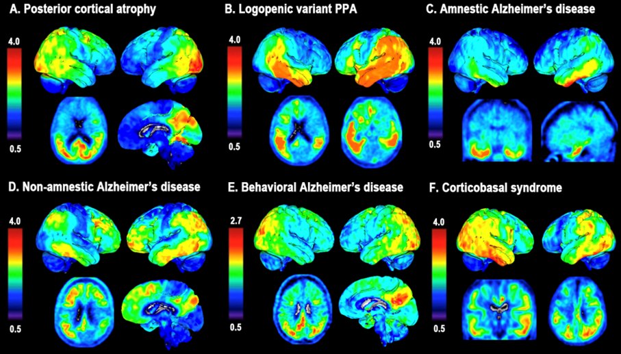
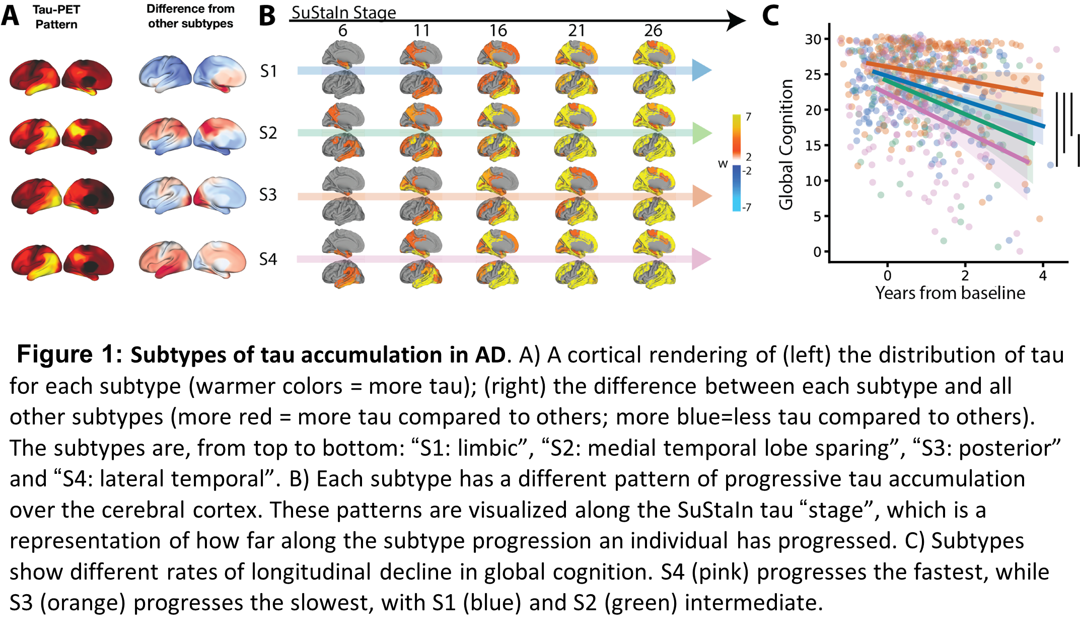
Background: The aggregation of tau pathology in Alzheimer’s disease (AD) is strongly associated with cognitive decline and local neurodegeneration. Tau pathology accumulates in the cerebral cortex in complex spatiotemporal patterns that differs across individuals, leading to diverse clinical phenotypes. Using positron emission tomography (PET), we recently characterized four subtypes of tau accumulation within the AD population, each with distinct clinicopathological profiles (Fig 1). Validation and further characterization of these subtypes is critical to better understand their clinical and potential therapeutic relevance.

 Methods: We applied the previously established Subtype and Stage Inference (SuStaIn) model to 1252 BioFINDER-2 (BF2) participants with available [18F]RO948 tau-PET. Linear models assessed subtype differences in CSF-biomarkers (corrected for Aβ40 and log-transformed). Linear mixed models were used to determine regional subtype-differences in atrophy (gray-matter volume) and cognition (MMSE and ADAS-cog delayed recall) over time. Models included age, sex, and baseline diagnosis, TIV (atrophy) and level of education (cognition) as covariates and were FDR-corrected.

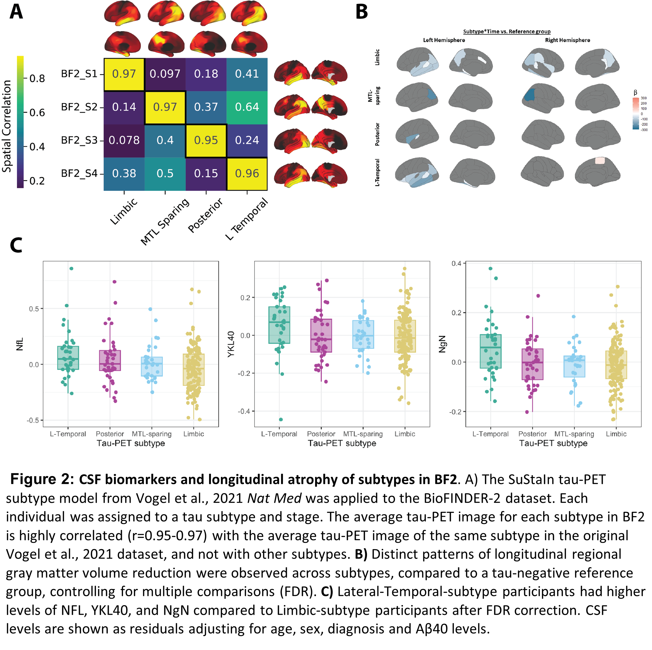
 Results: The four previously identified tau-PET subtypes were replicated in BF2 (Fig. 2A). Participants classified to the Limbic-subtype were older (F=33.3, p<0.01), while Posterior-subtype participants were less often APOE-ε4 carriers (χ2=119.83, p<0.001). Distinct patterns of longitudinal regional gray matter volume reduction were observed across subtypes (Fig. 2B). Posterior and Lateral-Temporal-subtype participants had higher levels of NFL, YKL40, and NgN compared to Limbic-subtype participants (pFDR<0.05, Fig. 2C). Limbic-subtype and Lateral-Temporal-subtype participants performed worse on ADAS-cog delayed recall (β=-1.00, pFDR<0.001) and MMSE (β=-1.01, pFDR<0.05) compared to the MTL-sparing and Limbic-subtype over time, respectively.

 Discussion: In a large single-site dataset, we confirm and extend findings that spatiotemporal subtypes of tau burden are differentiated by demographics and risk-factors. Cortical atrophy patterns are mostly consistent with the distribution of tau burden. The Lateral-Temporal-subtype showed higher tau burden and increased CSF markers of neurodegeneration, in line with the volume loss and cognitive decline.

Figures:

Authors & Affiliations:

Jacob Vogel1, Lyduine Collij2,3, Sophie Mastenbroek2,3, Alexa Pichet Binette3, Ruben Smith3,4, Sebastian Palmqvist3,4, Niklas Mattsson-Carlgren3,4, Olof Strandberg3,4, Rik Ossenkoppele3,5, Oskar Hansson3,4

1 Department of Clinical Science Malmö, SciLifeLab, Lund University, Lund Sweden

2 Amsterdam UMC, Department of Radiology and Nuclear Medicine, Amsterdam, the Netherlands

3 Clinical Memory Research Unit, Department of Clinical Sciences Malmö, Lund University, Lund, Sweden

4 Memory Clinic, Skåne University Hospital, Malmö, Sweden

5 Amsterdam UMC, Alzheimer Center Amsterdam, Department of Neurology, Amsterdam, The Netherlands

#6
Rik Ossenkoppele

AT(N) imaging in atypical variants of Alzheimer’s disease


Among patients with sporadic AD, the vast majority initially present with progressive decline of episodic memory function, followed by deficits in other cognitive domains. Testament to the great clinical variability of the disease, however, is the identification of several non-amnestic variants of AD, accounting for ~5-10% of all patients globally. These atypical variants include posterior cortical atrophy (PCA, the “visual variant of AD”), logopenic variant primary progressive aphasia (lvPPA, the “language variant of AD”), behavioral and/or dysexecutive variant(s) of AD and corticobasal syndrome (“motor variant of AD”). The common denominator across these non-amnestic variants of AD is that they often manifest at a relatively young age (<65 years at time of diagnosis) and demonstrate a lower prevalence of Apolipoprotein E [APOE] ε4, the major genetic risk allele for sporadic AD. Due to the relative paucity of age-related confounding co- pathologies, as well as the remarkable diversity of these syndromes, they provide an intriguing model to study mechanisms that drive clinico-anatomical heterogeneity in AD. This presentation will consist of three different parts. In part 1, I will demonstrate that amyloid positron emission tomography (PET) patterns are remarkably similar across AD phenotypes, while tau PET uptake and brain atrophy show a highly variant-specific distribution across the brain. In part 2, I will show that these findings generally translate to the typical (amnestic-predominant) spectrum of AD. In part 3, I will evaluate the evidence supporting a network-based spreading model of tau pathology and brain atrophy in atypical variants of AD (Figure 3). Altogether, AT(N) imaging in atypical variants of Alzheimer’s disease over the past decade has led to improvement of the diagnostic process and subsequent clinical management, and a better basic neuroscientific understanding of cognitive systems like vision, language and behavior.

Keywords: Atypical Alzheimer’s disease, tau, amyloid, atrophy, cognition

Figures:

FIGURE 1. Network spreading of tau pathology in atypical AD

(Adopted from Ossenkoppele et al. Brain, 2016)

 FIGURE 2. The clinical continuum of typical and atypical Alzheimer’s disease

(Adopted from Groot et al. NeuroImage Clinical, 2021)

FIGURE 3. Network spreading of tau pathology in atypical AD

(Adopted from Ossenkoppele et al. NeuroImage Clinical, 2019)

#7
Alexandre Bejanin

Neuroimaging of Alzheimer’s and vascular pathologies in Down syndrome 

Abstract: Individuals with Down Syndrome (DS) face an ultra-high risk of developing Alzheimer’s disease (AD) due to an extra copy of the APP gene on chromosome 21. This results in the nearly universal presence of AD neuropathological features by age 40 and a lifetime risk of dementia exceeding 90%. AD biomarkers in DS closely mirror the temporal pattern and pathophysiology seen in autosomal dominant AD, and there is as little variability in the mean age at symptom onset in either condition. Interestingly, the DS population is protected from some conventional vascular risk factors (e.g., hypertension) and rarely shows arteriolosclerosis; thus providing a unique model to understand the interaction between AD and vascular pathologies. Here, we compile novel evidence obtained from the Down Alzheimer Barcelona Neuroimaging Initiative (DABNI), one of the largest cohort of adults with DS with clinical assessments and multimodal biomarker, on the emergence of neurodegeneration and cerebral small vessel disease (SVD) lesions with AD. Using 3T-MRI and PET data, we characterized the topography and temporality of the pattern of atrophy and hypometabolism related to AD in DS. We further determined the prevalence and progression of SVD lesions (e.g., microbleed, white matter hyperintensities) in DS and define their associations with AD pathology and cognitive impairments. Our results highlighted a stereotypical pattern of neurodegeneration that strongly resembles the one of sporadic AD, even though some specificities are denoted. SVD lesions increased many years before AD symptom onset, and are closely linked with AD biomarkers, suggesting a direct connection to AD pathophysiology independent of vascular risks. Together, these results provide new insights into the interplay between AD and SVD and demonstrate that characterizing AD-related pathways in DS can also offer insights into AD pathophysiological processes.

Keywords: Genetically determined AD; Neurodegeneration; Vascular pathology; White matter hyperintensities; Microbleed.

Authors: Alexandre Bejanin1,2

1 Sant Pau Memory Unit, Department of Neurology, Hospital de la Santa Creu i Sant Pau, Biomedical Research Institute Sant Pau, Universitat Autònoma de Barcelona, Barcelona, Spain

2 Center of Biomedical Investigation Network for Neurodegenerative Diseases (CIBERNED), Madrid, Spain

#8
Gérard Bischof

18F-PI2620 Tau PET in AD and Non-AD
Neurodegenerative Diseases


Abstract: Second-generation tau PET tracers are currently being evaluated to visualize different tau isoforms. 18F-PI-2620 is a promising tau PET tracer as its affinity has been demonstrated in both clinical phenotypes of 4R and 3R/4R tauopathies. Several methodological approaches have been undertaken to improve the signal-to-background ratio, resulting in better quantification of 18F-PI-2620. In addition, several in vivo and ex vivo studies are now underway to evaluate the origin of the signal from 18F-PI-2620. Together, these studies are helping us to understand the tracer-binding relationship, but are also advancing our knowledge of the application of tau PET imaging in different disease phenotypes. This presentation will highlight the innovative ideas arising from the application of 18F-PI-2620 in the context of research specific questions, but also the advantages in the clinical implementation of 18F-PI-2620. Moreover, 18F-PI-2620 has been recently introduced in the context of novel disease phenotypes, which may improve our understanding of how different tauopathies produce different clinical phenotypes.  In addition, open questions regarding 18F-PI-2620 in the clinical context will be critically reviewed.


#9
Michela Plevani

Brain networks connectivity in AD and FTD


Abstract: Alzheimer’s disease (AD) and behavioural variant of frontotemporal dementia (bvFTD) are characterized by dysfunction of brain networks linked to specific cognitive and behavioural deficits. We present results of MRI-based studies investigating both commonalities and differences in brain networks connectivity in AD and bvFTD and how we used this knowledge to develop new strategies to modulate brain network dysfunction. Patients with a diagnosis of dementia (AD or bvFTD) and cognitively unimpaired older adults underwent 3T MRI imaging, including resting-state fMRI for functional connectivity assessment and diffusion weighted imaging (DWI) for structural connectivity analysis. Patients with AD and bvFTD showed convergent network-cognitive patterns in the default and salience networks, and divergent network-cognitive patterns in attention/executive networks, suggesting both compensatory and detrimental mechanisms. Structural connectivity patterns showed topographical differences between bvFTD and AD (anterior vs. posterior gradients) and a higher impairment in bvFTD, both at the cortical level and in tracts arising from the brainstem. Some connectivity alterations were already apparent in individuals at risk of dementia. Using electrical stimulation to modulate connectivity, we reported divergent effects on default mode network functional connectivity in AD based on the stimulation protocol. A protocol for targeted network modulation was developed based on individual resting-state fMRI data and is currently being tested in patients with AD or at risk for AD.

Keywords: Alzheimer’s disease, frontotemporal dementia, brain networks connectivity, MRI, non-invasive stimulation

Authors:

  • Michela Pievani, Laboratory Alzheimer’s Neuroimaging and Epidemiology, IRCCS Istituto Centro San Giovanni Di Dio Fatebenefratelli, Brescia, Italy
  • Giulia Quattrini, Laboratory Alzheimer’s Neuroimaging and Epidemiology, IRCCS Istituto Centro San Giovanni Di Dio Fatebenefratelli, Brescia, Italy
  • Lorenzo Pini, Padova Neuroscience Center, University of Padova, Padova, Italy
  • Marta Bortoletto, Neurophysiology Unit, IRCCS Istituto Centro San Giovanni di Dio Fatebenefratelli, Brescia, Italy
  • Debora Brignani, Department of Clinical and Experimental Sciences, University of Brescia, Brescia, Italy
  • Giovanni B. Frisoni, University Hospitals and University of Geneva, Geneva, Switzerland
  • Rosa Manenti, Neuropsychology Unit, IRCCS Istituto Centro San Giovanni di Dio Fatebenefratelli, Brescia, Italy
  • Francesca B. Pizzini, Department of Diagnostic and Public Health, Verona University Hospital, Verona University, Verona, Italy

NEWSLETTER

This website uses cookies to ensure audience measurement, as well as sharing or redirection to social networks. (Know more)
OK